Life 360 Physiotherapy Center, HIG-16, Sector 1, Shankar Nagar, Raipur, Chhattisgarh 492007
-
For Appointment: 09115719395 , 07000784644 , 07714908010
-

At Life 360 Physiotherapy Center, led by Dr. Nitu Verma, India’s first lady chiropractor, we are dedicated to delivering top-quality chiropractic and physiotherapy care. Recognized as the best chiropractor in India, Dr. Verma specializes in holistic treatments for pain relief, spinal alignment, and sports injury rehabilitation. Our clinic in Raipur provides personalized care designed to address the root causes of discomfort and restore balance to your body, helping you achieve long-term health and wellness. Experience relief and recovery with India’s leading chiropractic expert at Life 360 Physiotherapy Center.
For Appointment: 09115719395 , 07000784644 , 07714908010
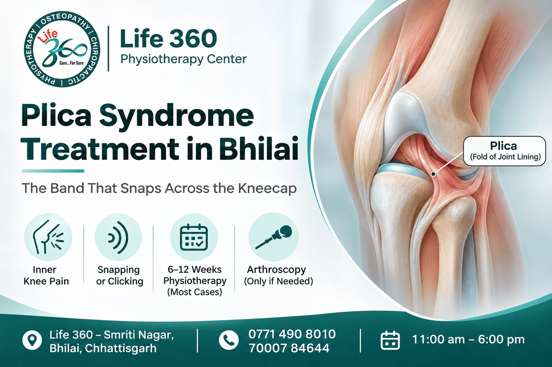
Quick answer: A plica is a leftover fold of joint lining inside the knee. When it thickens and gets pinched between the kneecap and the thigh bone, it causes inner knee pain, a snapping sensation, and pain after sitting with the knee bent. Most cases settle with six to twelve weeks of targeted physiotherapy, and arthroscopic resection is reserved for the few that do not.
She asked me to listen rather than look.
A 26-year-old data entry operator from Kumhari straightened her right leg slowly on the couch, and there it was: a soft deep click on the inner side, repeatable every time she passed about 40 degrees. Pain in that spot for seven months. Worse after the bus in from Kumhari with her knees jammed against the seat ahead, worse after two hours at her desk, better once she had walked for ten minutes.
Her MRI had been reported entirely normal, and she had been told the knee was fine.
I put two fingers on the inner edge of her kneecap and bent the knee slowly. A firm band rolled over the femoral condyle under my fingertips, and she said "that is it" before I asked anything.
Plicae are remnants of the membranes that divided the knee into compartments before birth. Most people have at least one and never know.
Suprapatellar plica, above the kneecap, sometimes nearly complete. Rarely symptomatic.
Mediopatellar plica, down the inner wall beside the kneecap. This one causes almost all the trouble, because it alone lies directly between kneecap and thigh bone.
Infrapatellar plica, the ligamentum mucosum, a cord running into the notch. Common at arthroscopy, almost never the source of pain.
Lateral plica, uncommon and usually silent.
So the practical statement is narrow: plica syndrome almost always means a thickened mediopatellar plica.
Repeated bending and straightening under load, or a bout of swelling, thickens the fold until it loses its softness and starts catching on the bone edge instead of sliding over it.
The sequence runs like this. Something irritates the knee, often a new activity or unaccustomed cycling. The joint produces extra fluid. The plica swells, turns fibrous, and each bend then drags a stiffened band across the condyle, which keeps the irritation going. The original cause disappears and the cycle continues on its own.
A direct blow to the inner knee can start it too, which is why I ask about handlebars and desk corners.
Pain sits on the inner side of the knee, one to two centimetres above the joint line, higher than where a meniscus hurts. Patients can usually cover it with a fingertip.
The timing pattern is the useful part:
Worst after the knee has been held bent a while: long bus or car rides, desk work, cinema seats
A sharp catch on the first few steps after getting up, which eases within a minute
Snapping or clicking at around 30 to 50 degrees as the knee straightens
Aggravated by cycling, stairs and repeated squatting
Often comfortable during a long walk on flat ground, which confuses people
Three structures sit within a few centimetres of each other and produce overlapping pain, so the location and the mechanics separate them.
The mediopatellar plica hurts above the joint line on the inner side, snaps in mid-range, settles with walking. A medial meniscus tear hurts at the joint line itself, pinches at the bottom of a deep squat and can lock the knee: meniscus tear treatment in Bhilai. Hoffa's fat pad hurts below the kneecap, dead centre, worst with the knee pushed into full extension in standing: Hoffa's fat pad impingement treatment in Bhilai.
Diffuse front-of-knee pain with no snapping band belongs with patellofemoral pain treatment in Bhilai.
Because MRI shows a plica reliably only when there is enough fluid in the joint to outline it, and because a visible plica is a normal finding that radiologists have no reason to flag.
Both halves of that matter. A dry knee hides the fold. A knee with a visible fold gets reported as normal anatomy, correctly, because most plicae are harmless. The diagnosis is clinical: a reproducible snapping band under the examiner's fingers, with pain the patient recognises as their own.
This is a diagnosis made with hands, not with film.
Palpation of the medial plica with the knee moved slowly from 90 to 0 degrees, feeling for a band rolling over the condyle.
Mediopatellar plica test. Knee bent to 90 degrees, kneecap pushed medially while the knee is extended, reproducing the pain and the snap.
The exact angle at which it snaps, in degrees, written down so we can compare it in six weeks.
Joint line tenderness and McMurray's, to argue against a meniscus.
Fat pad compression with the knee locked straight, to argue against Hoffa's.
Effusion, by sweep test, because a persistently wet knee means something else is driving it.
Hip abductor and quadriceps strength, both sides, recorded as numbers.
Sitting posture and seat height, including the bus and bike positions you spend most time in.
Thigh girth 15 cm above the kneecap, both legs.
Weeks 0–3, settle the irritation. Cut the positions that compress the fold: sustained deep bend, cross-legged sitting, hours with the knee flexed under a desk. An alarm to straighten the knee every 30 minutes. Soft tissue work along the medial retinaculum. Isometric quadriceps at 30 to 40 degrees, where the plica is not pinched.
Weeks 3–7, restore the mechanics. Hip abductor and external rotator strengthening, because a thigh rotating inward presses the plica harder against the condyle. Closed-chain quadriceps in a pain-free range. Kneecap mobilisation by hand. Cycling back in with the saddle raised so the knee never passes about 90 degrees.
Weeks 7–12, load it properly. Progressive squats, step-downs, single-leg work, and a deliberate return to the provoking position. By week ten the snap often remains while the pain has gone, which is an acceptable result.
Typical course: eight to twelve sessions across ten weeks, twice weekly at first. Most notice a change by week four. If nothing has shifted by week eight, I reconsider the diagnosis rather than extending the programme.
A single corticosteroid injection around the plica has a place when pain is severe enough to block any exercise, and it can also act as a diagnostic test. If a well-placed injection gives a few weeks of clear relief, that supports the diagnosis.
What it does not do is fix the problem. The fold is still thickened. I use the window the injection creates to get the loading programme done, and I would not repeat it more than once in a knee.
Do not stretch the knee hard into full bend to "free the band".
This is the advice I most often have to undo. Patients are given deep knee flexion stretches and heel-to-buttock work, which drags the thickened plica repeatedly across the bone edge, and they arrive worse than when they started. The plica does not need lengthening. It needs to stop being pinched while the muscles around it get stronger.
The second thing is the chair. Sitting through a nine-hour shift with the knee bent at 90 degrees and no interruption will keep this going no matter how good the exercises are. Raise the seat slightly, keep the foot forward rather than tucked back, and stand up every half hour. Those three changes do more than any machine in my clinic.
A knee locked short of full extension that will not release, suggesting a loose fragment rather than a plica
A hot, red, swollen knee with fever, which must be assessed the same day
A knee that keeps filling with fluid within days of being drained
Giving way with actual falls, rather than a catch that recovers immediately
Night pain at rest, weight loss, or pain that is worsening steadily month on month
Rapid swelling after an injury, which points to a ligament or cartilage injury instead
Is plica syndrome a real diagnosis, or a label for unexplained knee pain?
It is real, and it is also over-diagnosed. A thickened symptomatic mediopatellar plica exists and responds to treatment. It has also become a convenient name for front-of-knee pain nobody can explain. It should only be diagnosed when a snapping band is reproducibly felt and reproduces the patient's own pain.
Can a plica be seen on ultrasound?
Sometimes, in experienced hands, particularly a thickened fold with some joint fluid present. Dynamic ultrasound can occasionally show the band flicking over the condyle. It is operator-dependent, so I treat a negative scan as meaningless rather than reassuring.
If the plica is removed surgically, does it grow back?
The fold does not regrow, though scar tissue can form in the same region and a small number of patients stay symptomatic. Results from arthroscopic resection are generally good when the diagnosis was right, which is why the diagnosis matters more than the procedure.
Will quadriceps exercises make it worse?
The wrong ones will. Full-range leg extensions and deep squats early on compress the fold and flare it. Isometrics at 30 to 40 degrees and mid-range closed-chain work do not. Range matters more than load in the first month.
How long should I try physiotherapy before considering arthroscopy?
About three months of genuine, well-targeted treatment. If pain is unchanged and the band still snaps painfully, a surgical opinion is reasonable. Resection is a small procedure with a quick recovery, and still the second choice.
Does a knee sleeve help?
A thin sleeve helps some people by warming the joint and giving feedback, and does nothing for the fold itself. A few weeks is fine; permanent use is not, because compression comfort tends to replace the strengthening that changes things. Our Raipur page, plica syndrome treatment, covers the tests further.
Come in if you have inner knee pain above the joint line, if something snaps or clicks in mid-range every time you straighten the leg, if the knee is worst after long sitting and better after walking, or if your MRI came back normal and you were told nothing more could be found.
Life 360 — Smriti Nagar, Bhilai, Chhattisgarh
Phone: 0771 490 8010 · 70007 84644
Hours: 11:00 am – 6:00 pm
Email: [email protected]
Main centre: HIG-16, Sector 1, Shankar Nagar, Raipur 492007
Bring with you: any MRI or ultrasound reports, a note of how long you sit at a stretch on a working day, and shorts.
Dr. Nitu Verma (P.T.) is the founder and clinical director of Life 360 Physiotherapy Center, Shankar Nagar, Raipur, and of Life 360 at Smriti Nagar, Bhilai. She is widely recognised as India's first female chiropractor, and has more than fifteen years in clinical practice with over 50,000 patient consultations behind her.
Her training: Master of Physiotherapy (Sports) · Master Diploma in Chiropractic, Ackerman Institute, Sweden · Diploma in Osteopathy and DO Certification, Canada · Fellowship in Orthopaedic Manual Therapy (FOMT), Australia · Cranio-Sacral Therapy, USA · Pelvic Floor Osteopathy, Madrid School · Certified Pilates Instructor, Ireland.
She received the Nari Shakti Award 2026, and appeared on Doordarshan in a national discussion titled "Breaking the Myth About Chiropractic".
She treats spine and joint conditions non-surgically across Raipur, Bhilai, Durg and the wider Chhattisgarh region.
Clinics: HIG-16, Sector 1, Shankar Nagar, Raipur 492007 · Smriti Nagar, Bhilai
Appointments: 0771 490 8010 · 70007 84644 · [email protected]
Dr. Nitu Verma (P.T.) — Chiropractor and Physiotherapist, Life 360, Raipur and Bhilai.
General information only, and not a substitute for a hands-on knee examination.